The CDKL5 protein ......
What is CDKL5 ?
CDKL5 stands for Cyclin-Dependent Kinase-Like 5. A kinase is a protein that energises other proteins into action. In doing so, a kinase can also regulate cell function. There are about 500 kinases in the body. One particular group of kinases only become active when linked to another protein called Cyclin. They are therefore known as a Cyclin-Dependent Kinases (CDK) and they have an important role in the cell cycle whereby a single parent cell divides into 2 daughter cells.
The CDKL5 gene codes for a protein that is "Like" a Cyclin-Dependent Kinase in that it contains similar sequences of certain amino acids. CDKL5 does not however form a link with cyclin. There are 5 known Cyclin-Dependent Kinase-Like proteins. CDKL1-4 are also involved with brain development in some way, Although less seems to be known about them, there is some evidence that CDKL2 may share some functions with CDKL5.
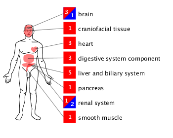
Studies have identified the presence of the CDKL5 protein in many different parts of the body. For children affected with CDKL5, the role of CDKL5 in the developing brain is obviously important, although in time, it may turn out that the function of CDKL5 in other areas may also be reflected in the clinical features....who knows.
An interesting fact about the CDKL5 gene is that its coding sequence is highly preserved and appears in many other species. This suggests that the protein has an important functional role that has been maintained through evolution.
The structure of the CDKL5 protein
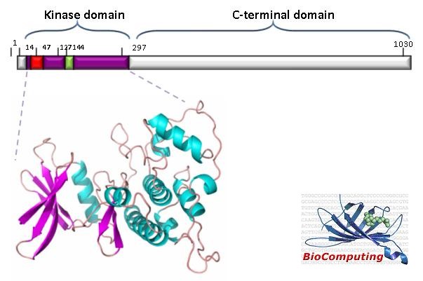
A protein consists of a chain of amino acids . There are 1030 amino acids in the CDKL5 protein which is divided into 2 regions, the catalytic or kinase domain, and the C-terminal domain.
Biochemical factors cause the protein chain to convolute into a 3-D structure. The function of the protein depends on its 3-D structure, which in turn depends on the correct sequencing of amino acids. Furthermore, although there is only one CDKL5 gene on the X-chromosome, there are at least 5 different forms of the protein that can result. One in particular that was previously identified, is thought to be the significant form in relation to CDKL5.
CDKL5 - the worker protein...
The Kinase domain is the active part of the protein in that, this is the part that energises other proteins into action. The kinase domain could be thought of as the "hands" of the protein that are doing the work.
The C-terminal is much longer in CDKL5 than it is in the other CDKL kinases. Initially, it was considered to be like the "legs" of the protein, in that research suggested that this part is involved in transporting the protein to the right area of the neuron. It may also have a role in regulating the function of the protein ... a sort of downing tools...! More research is needed to really understand its role in the overall function of the CDKL5 protein.
In terms of mutations, those that specifically affect the kinase domain will predominantly affect the kinase (hands) function of the protein so that it can get to work but just can't do its job properly or even do it at all. In contrast, a protein with a mutation affecting the C-terminal (legs) may still potentially be able to do its job but can't get to work to do it. There are some mutations of the kinase domain that will also produce a frameshift (deletions and insertions can do this) such that both the kinase and C-terminal domains are affected - so you end up with a protein with no hands or legs...

It is thought that CDKL5 is important both in the nucleus, where it probably interacts with MeCP2 - hence the association of CDKL5 with Rett Syndrome, and in the cytoplasm of the cell. To understand how mutations in CDKL5 might affect brain function it is worth knowing something about the development and structure of the brain.
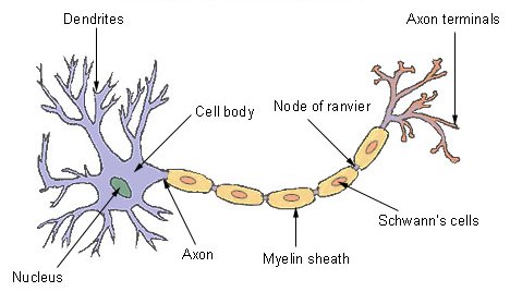
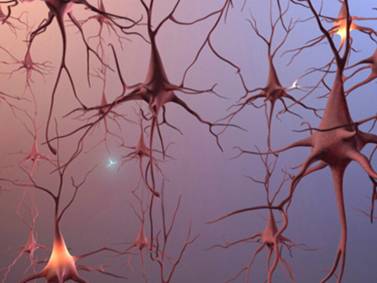
The structure of a neuron
The human brain contains about 86 billion neurons which are the main signal carrying cells. There are also other supporting cells called glial cells, such as astrocytes and oligodendrocytes, which actually constitute the majority of cells within the brain.
The structure of a neuron is dramatically different to a typical cell that you often see illustrated. The main cell body of a neuron has a network of branches (dendrites) that act like antennae, gathering information from other surrounding neurons.
Nerve cells generally function by carrying electrical signals. In fact, a “resting” neuron actually has a negative charge inside it. When stimulated by other neurons its negative charge can momentarily flip and become positive – an event called depolarisation. This positive charge can ultimately be carried down the axon of the neuron as an “action potential” and this forms the basis of the signal that the neuron then transmits to other nerve cells.
Courtesy of The Mind Project
Types of neuron
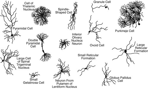
In the brain there are actually a whole variety of different types of neuron with very different appearances. However,broadly speaking they can be divided into 3 types according to their function, namely motor neurons that transmit motor information, sensory neurons for sensory information and interneurons which convey information between different types of neurons. They can also be classified as being excitatory (they cause their target neuron to fire off a signal) or inhibitory (inhibit neuron firing)
Alternatively, they can be classified according to the chemicals (neurotransmitters) that are used to transmit signals across synapses from one neuron to another. These include cholinergic neurons, dopaminergic neurons, GABAergic neurons, glutamatergic neurons and serotonergic neurons.
Glutamatergic neurons are commonly excitatory while GABAergic neurons are commonly inhibitory. This is relevant to CDKL5 as the anti-epileptic drug Vigabatrin (Sabril) acts by increasing the levels of GABA in the brain. Furthermore, studies on knockout mouse models have shown that behavioural changes can be mapped to GABAergic neurons in the forebrain in which the Cdkl5 protein has been removed.
Dendrites and spines
The numerous dendrites emanating from the cell body allow information to be gathered from far and wide. Signals are then passed down the axon to the axon terminal where information is passed on to other neurons across synapses (junctions between neurons).
The neurons are densely packed together within various layers of the brain, so you can start to imagine how this arrangement produces the hugely complex information gathering and processing network that is the brain.
(Illustration © 2012 Alzheimer's Association. www.alz.org All rights reserved. Illustration by Stacy Janis.)
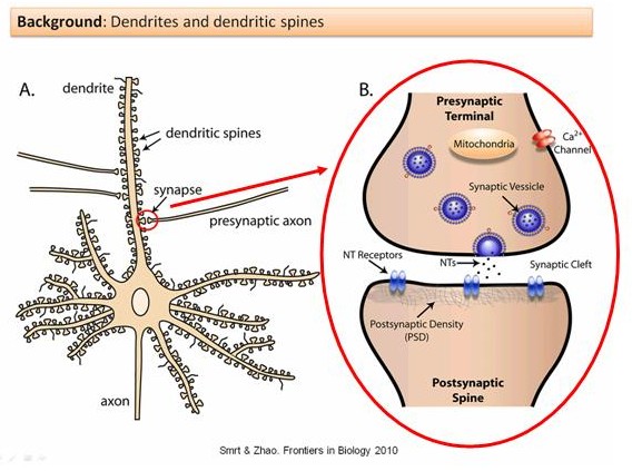
Dendrites themselves have small projections - known as spines. These are fairly "dynamic" structures, particularly in the developing brain, in that they can be seen to appear and disappear over relatively short periods of time. They are also able to change their morphology (shape or appearance) often within seconds.
This "plasticity" is a property largely determined, it seems, by active interactions from surrounding neurons - signals that are generated in the brain through the sensory input of our experiences. It is therefore thought that dendritic spines, their function and interactions, may also provide the basis of memory.
Research suggests that CDKL5 has an important role in their development and function. Lack of CDKL5 is associated with a reduction in dendritic arborization.
Early brain development
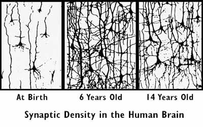
Previous studies on the normal development of the brain both before and after birth have shown how the morphology of neurons becomes increasingly complex. There is an increase in the size of neurons, and in the complexity of their dendrites (a process called arborization) which allows ever more connections to be made with other neurons around them.
This process is known as cell differentiation, and there are now a number studies investigating the role of CDKL5 in this process. Furthermore, as more studies are published, the complexity of the controlling pathways, as you might expect, are becoming more evident.
Modified from Amendola et al. "Mapping Pathological Phenotypes in a Mouse Model of CDKL5 Disorder." PloS one 9.5 (2014): e91613.
So, what happens in CDKL5?
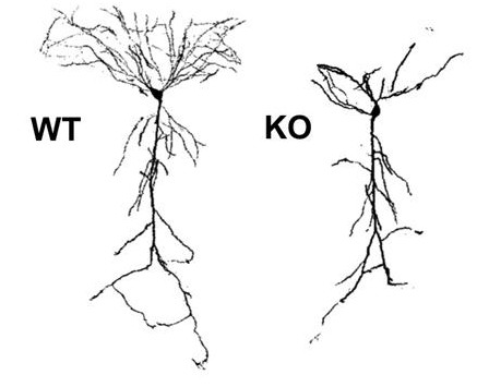
As studies on mouse models become available, we are learning more about what happens to developing neurons when the CDKL5 protein is deficient. In the absence of the CDKL5 protein, neurons appear to be underdeveloped compared to normal neuron development.
The adjacent figure shows a developing neuron in a normal mouse - usually referred to as a wild-type (WT) mouse in scientific publications - compared to that in a CDKL5 knockout (KO) mouse, in which the CDKL5 protein is absent. The WT mouse neuron is developing a normal complex pattern of dendrite branching (arborization) whereas the KO mouse neuron is much less branched and developed.
Remembering that dendrites and spines gather signals (information) from surrounding neurons so that they can be passed on to other neurons, you can start to understand how the function of these underdeveloped neurons will be impaired. The consequential clinical features and symptoms that are produced will obviously depend on where in the brain these affected neurons are.
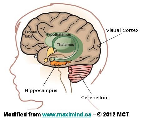
CDKL5 in the brain
The CDKL5 protein is probably distributed throughout all the neurons of the brain. However, there is evidence for the activity of the CDKL5 protein in specific areas of the brain. We know that children with CDKL5 often have visual cortical impairment, suggesting specific involvement of the visual cortex.
Most children with CDKL5 do not walk - Ellie doesn’t although she has normal power in her legs and arms - I know, I've been given a good kick... or a brilliant "hand off" on many an occasion...! However, she also has relatively poor balance and co-ordination which might be related to her poor vision or possibly to involvement of her cerebellum - a particular part of the brain responsible for co-ordination.
Studies on the activity of CDKL5 have also focused on an area of the brain called the hippocampus - involved in learning and memory. This is because the neurons here only really start to develop after birth and so the influence of the CDKL5 protein can more readily be studied.
So far, I have tried to provide an overview of the CDKL5 protein in terms of its structure and the effect that CDKL5 mutations have on the brain. That is, we are starting to understand the effects of CDKL5 mutations on the basic structure of neurons. However, to really understand the effects that CDKL5 mutations have on function, it is necessary to determine, study and understand the various molecular pathways in which the CDKL5 protein is involved ...... and this is where it really starts to get technical - hence a picture.....!
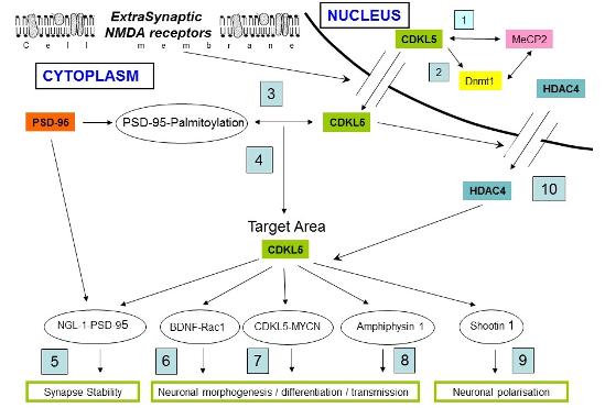
1. CDKL5 is a brain MeCP2 target gene regulated by DNA methylation - Carouge et al.
2. Cyclin-dependent kinase-like 5 binds and phosphorylates DNA methyltransferase 1 - Kameshita et al
3. ExtrasynapticN-Methyl-D-aspartate (NMDA) Receptor Stimulation Induces Cytoplasmic Translocation of the CDKL5 Kinase and Its
Proteasomal Degradation - Rusconi et al.
4. Palmitoylation-dependent CDKL5–PSD-95 interaction regulates synaptic targeting of CDKL5 and dendritic spine development - Zhu et al.
5. CDKL5 ensures excitatory synapse stability by reinforcing NGL-1–PSD95 interaction in the postsynaptic compartment and is impaired in
patient iPSC-derived neurons - Ricciardi et al.
6. CDKL5, a Protein Associated with Rett Syndrome, Regulates Neuronal Morphogenesis via Rac1 Signaling - Chen et al.
7. CDKL5, a novel MYCN-repressed gene, blocks cell cycle and promotes differentiation of neuronal cells - Valli et al.
8. Identification of amphiphysin 1 as an endogenous substrate for CDKL5, a protein kinase associated with X-linked neurodevelopmental
disorder - Sekiguchi et al.
9. CDKL5 and Shootin 1 interact and concur in regulating neuronal polorisation - Nawaz et al.
10. HDAC4: A key factor in brain development alterations in CDKL5 Disorder - Trazzi et al.
Since I produced this diagram, more research has been undertaken identifying more substrates and pathways that CDKL5 is involved in...see research updates.
Future Research
As time goes by, further research will demonstrate the involvement of CDKL5 in other molecular pathways. Two particular research tools to study the function of CDKL5 at the molecular level have been developed, namely iPS technology and the development of knockout mouse models. Mouse models also allow the study of some of the clinical aspects. There is research into protein replacement therapy (PRT), as well as into the use of genetic engineering such as CRISPR . An excellent review of the biology of CDKL5 was published in 2019 and is available on-line.
iPS cells - induced pluripotent stem cells, are cells that have the ability to grow and divide into any specialised cell, like a muscle cell, a bone cell or a nerve cell for the study of CDKL5. The clever bit is that by using technology that was first described in 2006, iPS cells can now be “made” from ordinary cells like skin cells.
By taking a sample of skin cells from a child with a given CDKL5 mutation, it is possible to turn these into iPS cells which then develop into neurons that have the same mutation. These can then be studied and compared to neurons with a normal CDKL5 gene.
Although there are limitations to this technique, it allows for a detailed study of the function of the CDKL5 protein in the neuron, and also for the development of possible drug treatments. The scientists who developed these techniques were awarded the Nobel prize for medicine in 2012.
A knockout mouse is one where a specific gene has been either removed from the mouse DNA or just disrupted. This then allows for the study of many characteristics of the mouse when the specific protein coded by that gene is absent.
A number of knockout mouse models have been established for CDKL5.
You can also have a "knockin mouse" where the mouse has a particular mutation or other specific protein coding sequence inserted into its DNA. This has been developed for the R59X mutation in exon 5 which a number of children with CDKL5 have.